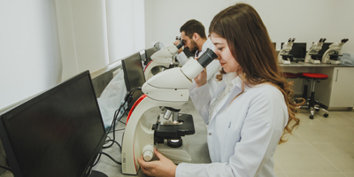
Qualified Personnel are being trained at EUL Pathology Laboratory

European University of Lefke (EUL) Vocational School of Health Services Academician Prof. Dr. Tahir Ercan Patıroğlu gave information about pathology laboratory which is established within the body of EUL and explained pathology.
Patıroğlu: Pathology performs an important role in diagnosis and treatment
Patıroğlu explained pathology as a science of diseases which performs an important role in diagnosis and treatment through examining organ-tissue biopsies, body fluids and cell specimens to determine structural and functional changes in cells, tissues and organs using different morphological, immunological and molecular methods, investigating the underlying causes, causes and symptoms of disease in patients.
Patıroğlu said that “Pathology sheds light on the direction of the treatment of the disease by giving the name of the disease (diagnosis) and giving additional information about the clinical course of the disease”. Patıroğlu said that pathology laboratories should have a pathologist, pathology technicians, laboratory equipment, consumables and microscopes.
Applied Educational Opportunity in Equipped Pathology Laboratory
Patıroğlu said that the Department of Pathology Laboratory Techniques educates qualified technicians who prepare body tissues or fluids for microscopic examination, perform necessary administrative, technical preparations and supervision before, during and after examination, identify and solve problems, and use eye and hand skills well and underlined that the students receive applied trainings in equipped pathology laboratory.
Patıroğlu stated that pathology laboratory which is established within the body of European University of Lefke, students of Pathology Laboratory Techniques Department use carousel-type semi-closed, fully automated tissue monitoring device to make various chemical solutions, and in the last stages, they do tissue follow ups in paraffin. Patıroğlu stated that the students gets tissue blocks by embedding the tissue pieces they extracted from the tissue tracking device into paraffin molds with the tissue embedding device and added that students made sections of 4-6 microns in thickness using rotary microtome from the tissue blocks obtained. Patıroğlu emphasized that the cuts are taken from the hot water pool on the slide and then stained with Hematoxylin & Eosin and by doing so they are prepared to be delivered to the relevant Pathology Expert for microscopic evaluation.
